|
Data Import
Data import pages. a)Āimport of tabular dual channel unnormalized data. Below the heading of the columns drop-down boxes are used to provide information for import. b)ĀMicroarray import view. The table on the left side can be used to select microarrays by name, the right side shows the scanned microarray image and data characteristics. The value distribution of all arrays is given in black, the selected array is marked in red. c)ĀNormalization parameter selection page d)Āoverview of imported and annotated microarray expression data. Probes are shown in the rows, the columns show gene information and sample expression.
|
|
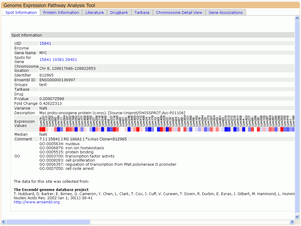
Gene Information
Gene information pages. a)ĀShows an overview of probe 15841 that measures expression level of the MYC gene. The information shown can be modularly extended. b)Āassociated genes for this probe, overlaid with differential gene expression results c)Āshows literature references and a short description derived from RefSeq, d)Āshows protein information for the gene. The upper part shows the coding regions, the lower part shows features for the different transcripts.
|
|
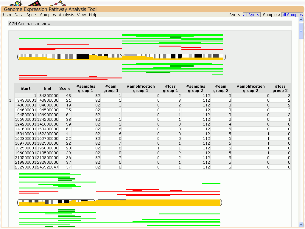
Data Analysis
Data Analysis result views. Results are shown for activated B-cell (ABC) type cancer samples and germinal center B-cell (GCB) type cancer samples: a)ĀM/A plot of moderate t-test result comparing ABC with GCB b)Āhierarchical clustering results. The color of the samples marks the different disease types. c)ĀPCA analysis results. Characteristical probes for disease were used as source for clustering. d)ĀCGH profile comparison. The yellow bar in the chromosome shows the difference between the profiles. Above the chromosome the CGH Profile of the ABC group is shown, the GCB group is shown below.
|
|
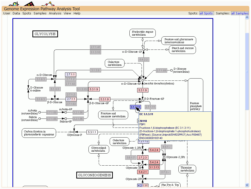
Data Interpretation
Views of data interpretation. The overlaid differential expression values are the result of the t-test shown in data analysis. Node colors reflect the differential gene expression. The light gray nodes represent associated genes not on the array. a)ĀComparing the genes with the lowest p-value shows enriched lymphocyte activation in the GO Term enrichment analysis. b)ĀGlucolysis KEGG map overlaid with differential expression result. c)Ā gene association network of the glucolysis genes. d)ĀChromosomal location detail view of a chromosome part containing a differential expressed gene. Genes measured on the chip are marked in yellow on the chromosome, differential gene expression is shown above and below the chromosome.
|